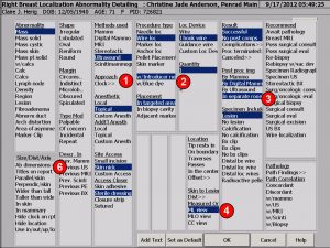
(1) Indicate the “approach” by clock face reference in report, lesion to lesion.
(2) Surgeon can now specify that methylene blue dye was injected.
(3) Users can now specify that post localization imaging was performed in a separate room as a separate procedure to capture additional reimbursement when multiple imaging modalities are used.
(4) Added skin to lesion measurement option and the ability to specify the radiographic view name.

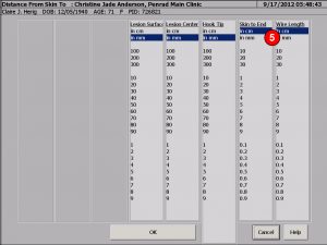
(5) Specify the length of the wire and length protruding from the skin to verify location prior to excisional procedure.
Finding example: “A 20 cm J-hook wire was inserted into the targeted area through an introducer device under ultrasound guidance”.
Impression example: “Wire localization for the lesion in the right breast at 5 o’clock posterior depth 8 cm from the skin to the surface of lesion and 9 cm from the skin to the geometric center of lesion measured on the ML view was successful with the end protruding 12 cm.”
If bracketing lesion, just indicate more than one wire and select “On boundary”. This will generate for example: “Post placement digital mammographic imaging demonstrates the tips demarcate the boundaries of the lesion”.
(6) As in all detailed exams the ability exists to indicate 3 dimensions of orientation, distance from skin, chest and nipple of the abnormality by tapping the “Ab dimensions” selector in the Size/Dist/Axis window and selecting values.
Added a Preview report button on bottom of main exam screen for instant report preview.
Recommendation for product development?
[email protected] | 763.475.3388
© 2019 PenRad Technologies, Inc.
