Ultrasound additions
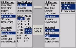
To facilitate enhanced ultrasound reimbursement, discrete scanned regions were added to differentiate exams as Complete or Limited. Also a series of requested descriptors and new US BI-RADS modifiers were added.

Added Supraclavicular as a region scanned.
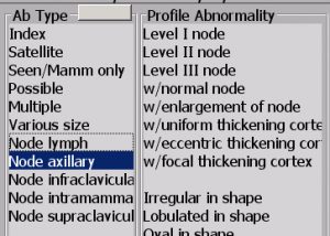
Added ability to specify axillary lymph node location as level 1-3.
Added ability to independently specify nodes as normal or enlarged.
Expanded cortex thickening to include; uniform, eccentric or focal.
Added ability to specify nodes with or without extranodal extension.
Added descriptor “not circumscribed margin”.
Added descriptor “without posterior features”.
Added ability to specify an abnormality as the “index” or “satellite”, and if satellite, the distance from index lesion.
When scanned regions are indicated as complete, the exam title incorporates “Complete”. Automatically following the referenced comparisons statement, the phrase “The four quadrants and retroareolar regions were scanned.” is inserted to differentiate the exam as complete for billing. Exams indicated as “Limited” reflect “Limited” in exam title, and the specific regions scanned are automatically incorporated into the report.
Exam titles automatically include various region specifiers. For example; “and axilla”, “axilla only”, “targeted”, and “supraclavicular”. These regions scanned are also automatically reflected in the body of the report.
Independently the retroareolar (R/A), axilla, and supraclavicular can be included in the region scanned. When bilateral scanning methods and regions scanned are alike a common bilateral scanned phrase is created.

To facilitate the callout of regions scanned within the report body, the option is provided by breast to reflect “the clock(s)”, “the quadrants”, “the axilla”, “the retroareolar”, and “the supraclavicular”. Scanned region phrase logic offers automatic consecutive region phrase consolidation for example; upper inner (UI) and lower outer (LO) reflects; outer aspect of the breast. For individual clock regions call out, if 11, 12, 1, 2, 7, 8 were selected, the scanned region phrase outputs as; 11-2 o’clock and 7-8 o’clock regions. When scanning methods and regions scanned are alike for bilateral exams, a common scanned “both” statement is created.
For automation, the technologist ultrasound imaging screen collects area scanned and technique used replacing the “ultrasound worksheet”. This data automatically populates the narrative report for the radiologist.
Mammogram and MRI additions
Added to Mammogram and MRI the ability to optionally specify an abnormality as the “index” or “satellite” lesion, and if satellite, the distance from the index lesion.
MRI FYI: Frequently PenRad is told “you’re missing non-mass like enhancement” on the MRI detail screen. Non-mass enhancement is available as a descriptor from the “Area” category (it is not mass like).
Recommendation for product development?
[email protected] | 763.475.3388
© 2019 PenRad Technologies, Inc. 730-23-00356 AB
